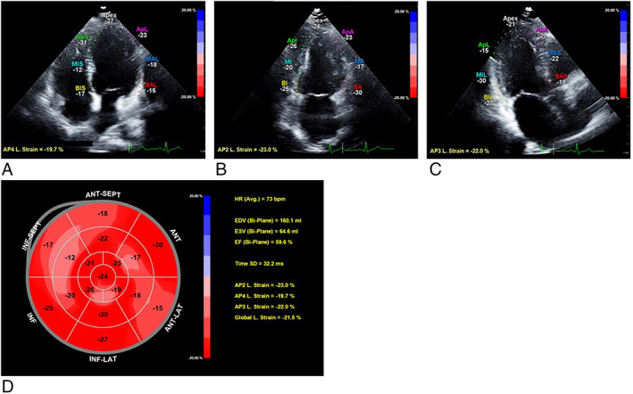
Figure 1.

Measurement of global longitudinal strain (GLS) by speckle‐tracking analysis in an obesity patient [45‐year‐old woman, body mass index (BMI) 38.4 kg/m2]. (A) Apical four‐chamber view with measurement of longitudinal strain. (B) Apical two‐chamber view with measurement of longitudinal strain. (C) Apical three‐chamber view with measurement of longitudinal strain. (D) Bull's eye graph showing longitudinal strain for all myocardial segments, of which a weighted mean was used to derive GLS.
