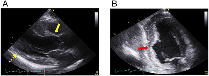
Figure 2.

(A) Parasternal long‐axis echocardiographic view at end‐diastole showing left ventricular wall thinning in the basal anteroseptal (yellow arrow) and mid‐inferoseptal (yellow dotted arrow) walls. (B) Apical two‐chamber echocardiographic view showing an aneurysm from the mid‐inferoseptum to the posterior walls (red arrow).
