
Figure 3.
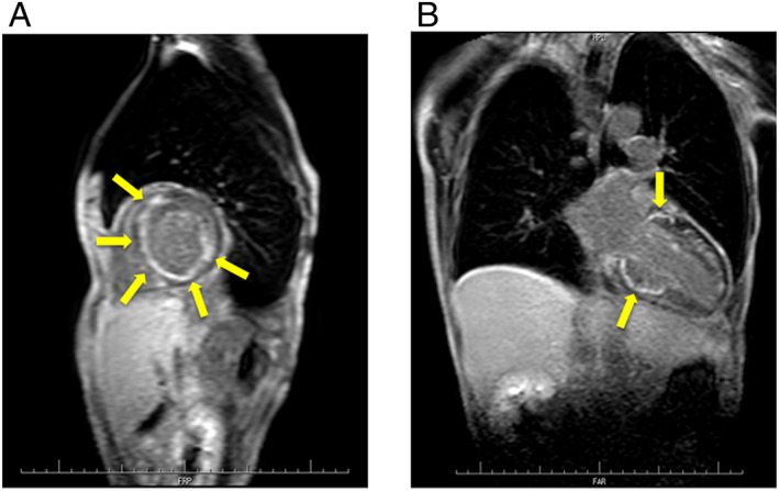
(A) Cardiac magnetic resonance (CMR) in the short‐axis view showing delayed gadolinium enhancement in the middle layers of left ventricular (LV) anteroseptal, inferoseptal, posterior, and lateral walls (yellow arrows). (B) CMR in the long‐axis view showing delayed gadolinium enhancement in the middle layers of LV anteroseptal, inferoseptal, and posterior walls (yellow arrows).