
Figure 6.
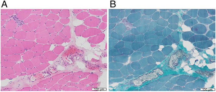
Muscle biopsy of the left biceps brachii showing fibre size variation, sporadic necrotic fibres, and few inflammatory cell infiltration (A, haematoxylin–eosin stain; B, modified Gomori Trichrome Stain, dimensional bar = 100 μm).
Official websites use .gov
A
.gov website belongs to an official
government organization in the United States.
Secure .gov websites use HTTPS
A lock (
) or https:// means you've safely
connected to the .gov website. Share sensitive
information only on official, secure websites.
Muscle biopsy of the left biceps brachii showing fibre size variation, sporadic necrotic fibres, and few inflammatory cell infiltration (A, haematoxylin–eosin stain; B, modified Gomori Trichrome Stain, dimensional bar = 100 μm).