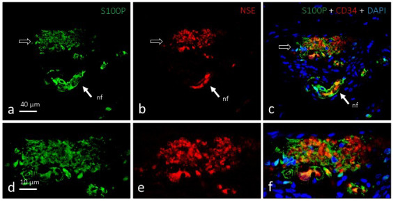
Figure 3.

Dual immunofluorescence for S100 protein (a and d; green fluorescence) and neuron‐specific enolase (b and e; red fluorescence) in a single Ruffini's corpuscle (arrows). The axonal branch and glial cells exhibit complementary localisation (c and f), are intimately linked, and occupy most of the corpuscle. nf, nerve fibre supplying the corpuscle. Objective: 63×/1.40 oil; pinhole: 1.37; XY resolution: 139.4 nm; and Z resolution: 235.8 nm
