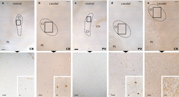
Figure 3.

Immunohistochemical staining of the sheep Cl. Immunoperoxidase reaction shows the distribution of CB in the rostral (a) and caudal (b) parts, PV in the rostral (c) and caudal (d) parts, and CR caudal part (e) immunoreactivity in the Cl (dashed line). Below (a), very few and weakly stained CB‐ir cells are spotted. Higher magnification of the frames below (b) displays CB‐ir cells and scarce fibers (enlarged in the inset). Below (c) are rare fibers with no real soma stain for PV. Below (d), higher magnification shows a moderate density of PV‐ir fibers and two positive neurons (enlarged in the inset). Below (e), higher magnification shows a dense network of CR‐positive fibers (enlarged in the inset). Cl, claustrum; ctx, cortex; Pt, putamen. Scale bars =500 μm (upper row), 100 μm (lower row), 10 μm (insets)
