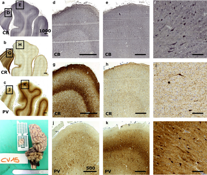
Figure 4.

Immunocytochemical staining of the sheep V1. Immunocytochemistry reveals the patterns of calcium‐binding proteins in V1 (e, h, k) and the peristriate area (d, g, j). Calbindin is found in most of the cortical thickness (a, d, e, f), staining round interneurons and some pyramidal cells, while calretinin is much more localized, notably in layer 1 (b, g, h, i). Parvalbumin shows also a large positivity throughout the cortical thickness (c, j, k, l) with a marked neuropil band in V1 (k). Positive cells are rather large and round multipolar, with weaker staining of some pyramidal neurons (l). Bar in (a), (b), (c) is 1000 µm; bar in (d), (e), (g), (h), (j), and (k) is 500 µm, (f), (i), (l) bar is 50 µm
