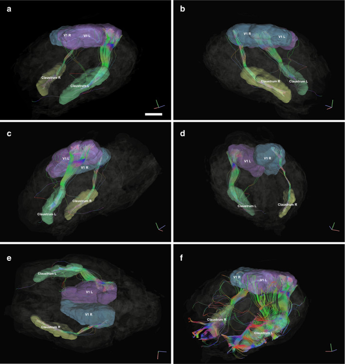
Figure 5.

Representation of fiber tracts running between the left and right Cl (yellow and red, respectively) and the left and right visual areas (in blue and green, respectively). MRI images represent the orthogonal planes. (a) Left antero‐lateral view. Indicative bar = 1 cm. (b) Right antero‐lateral view. In (a) and (b), the extent of the tracts in the claustra can be appreciated, as well as the posterior access of the fibers, originating from the occipital aspect of the visual cortex. (c) Left Lateral view. (d) Right postero‐lateral view. (e) Ventral view where the left tracts can be seen reaching up to the visual cortex in the dorso‐occipital cortex. (f) Left antero‐lateral view, where the Cl is used as a seed to map the tracts originating from it. The fibers exiting the Cl are very rich. Note the projection to the anterior part of the visual territory, emerging from the caudo‐dorsal part of the Cl
