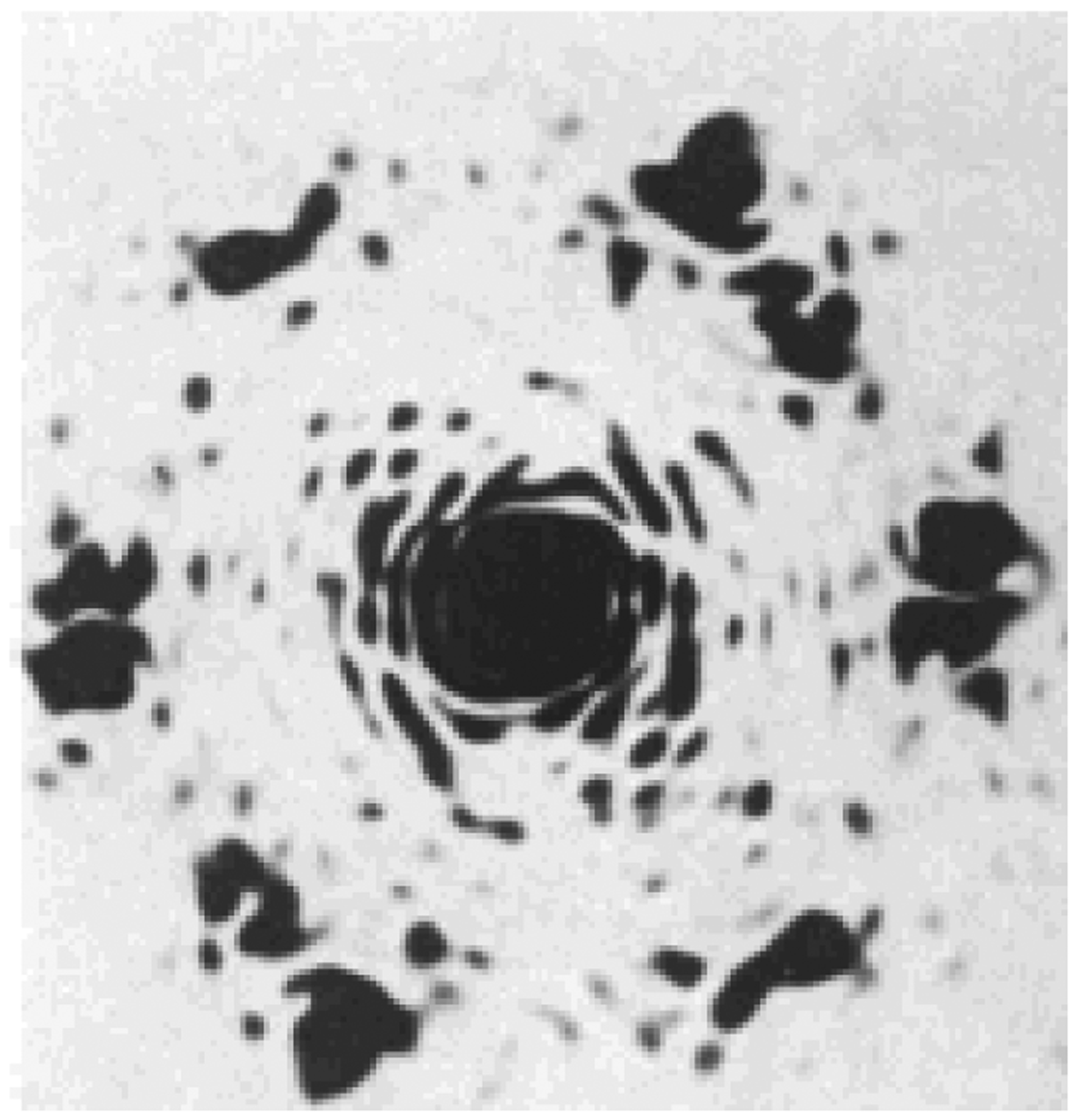
Fig. (1).

Optical diffraction patterns from the electron micrographs showing the chitin crystallites in exocuticle from Oryctes rhinoceros, pharate adult femur. Six hexagonally arranged spots may be seen showing the near-perfect hexagonal packing of the chitin crystallites, A. C. Neville, et al. (1976) [1].
