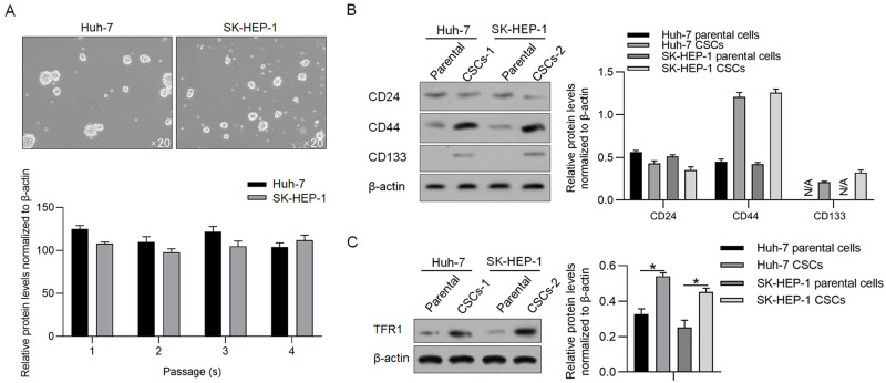
Fig 2. TFR1 protein levels in CSCs derived from hepatocarcinoma cell lines.
A. Spheres were observed by culturing Huh-7 and SK-HEP-1 cells in serum-free medium. Images were obtained using a 20× objective. B. Stemness markers, including CD24, CD133 and CD44, were measured by performing western blotting. C. TFR1 protein expression was measured in CSCs and compared with that in the parental cells. *P<0.05, vs. parental cells.

