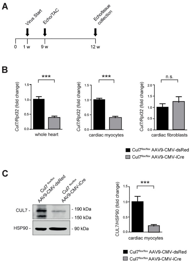
Fig 1. Generation and validation of cardiomyocyte-specific Cul7-/- mice.

Experimental strategy and timeline (A). On day 4 to 5 after birth mice were transduced pericardially with AAV9-CMV-iCre or AAV9-CMV-dsRed serving as control group. At week 9 mice underwent sham or TAC surgery und were euthanized for further analysis at week 12. (B) Realtime-PCR of Cul7 mRNA in whole heart lysates (WH, left panel, n = 7–9 mice/group), isolated cardiac myocytes (CM, middle panel, n = 7 mice/group) and isolated cardiac fibroblasts (CF, right panel, n = 7–9 mice/group). (C) Immunoblot analysis for CUL7 protein in isolated CM (left panel) and quantification thereof (right panel). n = 3–7 mice/group. Data are shown as fold change normalized to controls and expressed as mean ± SEM. ***p<0.001 (unpaired t-test). WH: whole heart, CM: cardiac myocytes, CF: cardiac fibroblasts.
