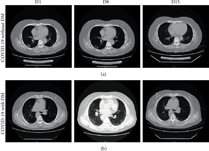
Figure 1.

Representative chest computed tomography (CT) images of COVID-19 pneumonia in a nondiabetes mellitus (DM) case and a DM case. (a) A 54-year-old woman with COVID-19, but not DM: chest CT images showed ground-glass opacity (GGO) and patchy consolidation with peripheral and subpleural distribution, which had been absorbed at 15 days after hospitalization with treatment. (b) A 58-year-old woman with both COVID-19 and DM: chest CT images showed bilateral multiple lobules and subpleural diffuse consolidation. Chest CT showed that there was still a small amount of bilateral ground-glass opacity at 15 days after hospitalization with treatment.
