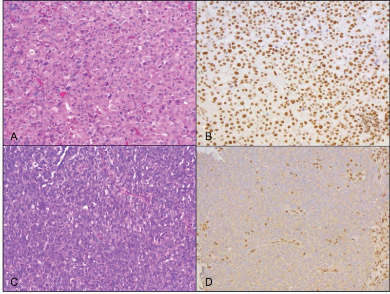
Fig. 2.

Histology of one SR-SNUC case and one SD-SNUC case. Hematoxylin and eosin (H&E) stained section of the SR-SNUC case demonstrates squamoid morphology characterized by tumor cells with abundant eosinophilic cytoplasm and distinct cell borders (A, ×20); IHC for SMARCB1 shows strong nuclear staining in the tumor cells (B, ×20). The SD-SNUC case shows basaloid morphology characterized by tumor cells with high nuclear to cytoplasmic ratio (C, H&E, ×20); SMARCB1 nuclear expression is lost in the tumor cells and retained in the background nonneoplastic inflammatory and stromal cells (D, ×20). SD-SNUC, SMARCB1 -deficient sinonasal undifferentiated carcinoma; SR-SNUC, SMARCB1 -retained SNUC.
