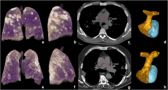
Fig. 4.
Exemplifying cases: a survivor (top) and non-survivor (bottom) patients. CT 3D volume rendering of lungs in antero-posterior view (a, e) and lateral view (b, f), CT axial images with mediastinal window (c, g) for main pulmonary artery diameter (MPAD) measurement (red lines in c, g), and 3D volumetric reconstruction of pulmonary arteries (d, h) in a survivor (top) and a non-survivor (bottom) COVID-19 patient, both males of 74 and 76 years, respectively, both with history of hypertension and treated ischemic cardiomyopathy, both suffering from fever (> 37.5 °C) and caught from 7 days. At the admission, WBC/mm3 were 4.36 and 3.8, CRP 0.93 and 1.1 mg/dL, LDH 295 and 378 U/L, and creatinine 0.93 and 1.1 mg/dL in survivor and non-survivor, respectively. The oxygen saturation was 90% and 94% for survivor and non-survivor, respectively, with higher well-aerated lung volume in the survivor patient (3882 mL vs 2382 mL, violet parenchyma on 3D lung volume rendering in a, b, e, f) with pneumonia (bright parenchyma in a, b, e, f), involving from 25 to 50% of lung volume in both cases. Pulmonary artery diameter was normal (27 mm) in the survivor (c, d) while it was enlarged (32 mm) in the non-survivor (g, h) who died 9 days after hospital admission

