


Figure 5.
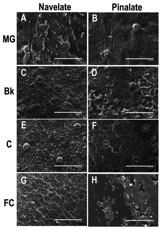
Epicuticular wax morphology during fruit ripening. Scan electron micrographs of Navelate (A) and Pinalate (B) mature green, Navelate (C) and Pinalate (D) breaker, Navelate (E) and Pinalate (F) colored, and Navelate (G) and Pinalate (H) full-colored fruit ripening stages. The same scale was used in all the micrographs. Scale bars: 100 μm.