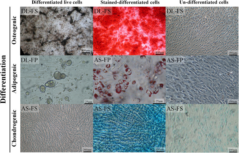
FIGURE 3.
SMSCs derived from different synovial tissue sources in DL and AS breeds successfully differentiated into adipocytes, osteocytes, and chondrocytes under optimal conditions. In the late stages of osteogenesis after culturing in an osteogenic medium for 28 days, calcium deposits were revealed as brown–black lines or spots under a 4× objective phase-contrast microscope and as red-brown after being stained with alizarin red. The lipid-vacuole and lipid-droplet formation of adipocytes was observable at 4 days of adipogenic differentiation; the cells were fixed and stained with Oil Red O to identify the lipid vacuoles (red). For chondrogenic differentiation, the cells were cultivated in a chondrogenic medium for 35 days; sulfated proteoglycans, including hyaluronic acid, were then stained with alcian blue 8GX (bluish-green/blue). The control cultured un-differentiated cells had a fibroblastic morphology under phase contrast similar to that observed before differentiation to chondrocytes, adipocytes, and osteocytes.

