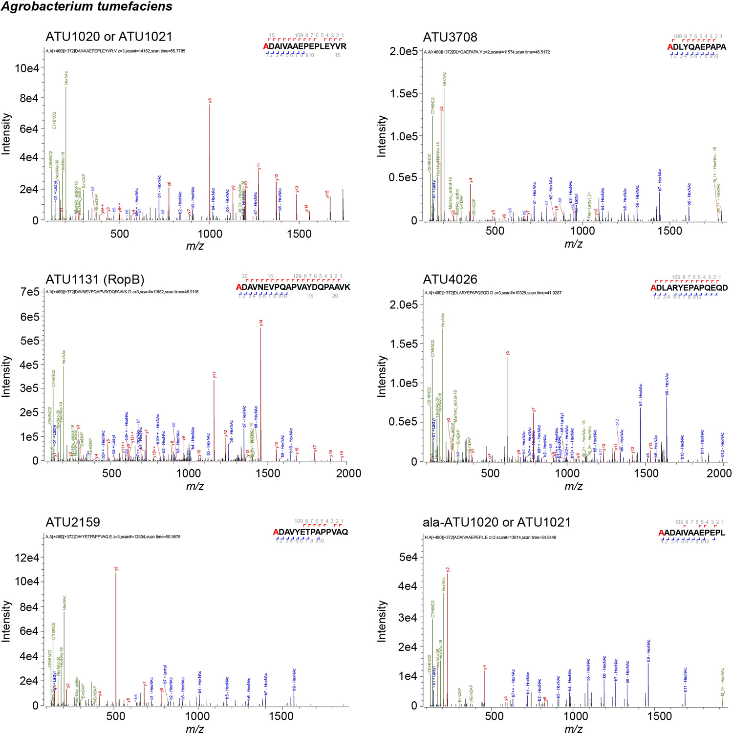
Extended Data Fig. 3. Automated spectrum assignment identifies PG modifications on β-barrel proteins from A. tumefaciens.

Representative MS/MS spectra for A. tumefaciens β-barrel proteins covalently attached to mDAP (m) residues of PG. Residues with PG tripeptide modification are highlighted in red. Spectra are shown as annotated by Byonic using an unbiased search approach.
