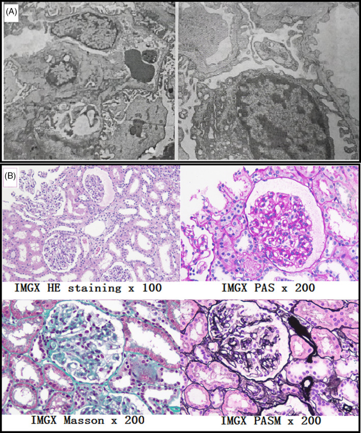
Figure 1.

Renal pathological examination results of case IV‐1. Mild proliferation of mesangial cells and a thin glomerular basement membrane (less than 200 nm) were observed in the pathological examination of kidneys (A). Furthermore, IgA, IgG, and C1q tests using immunofluorescence staining of glomerular vascular wall were positive (B)
