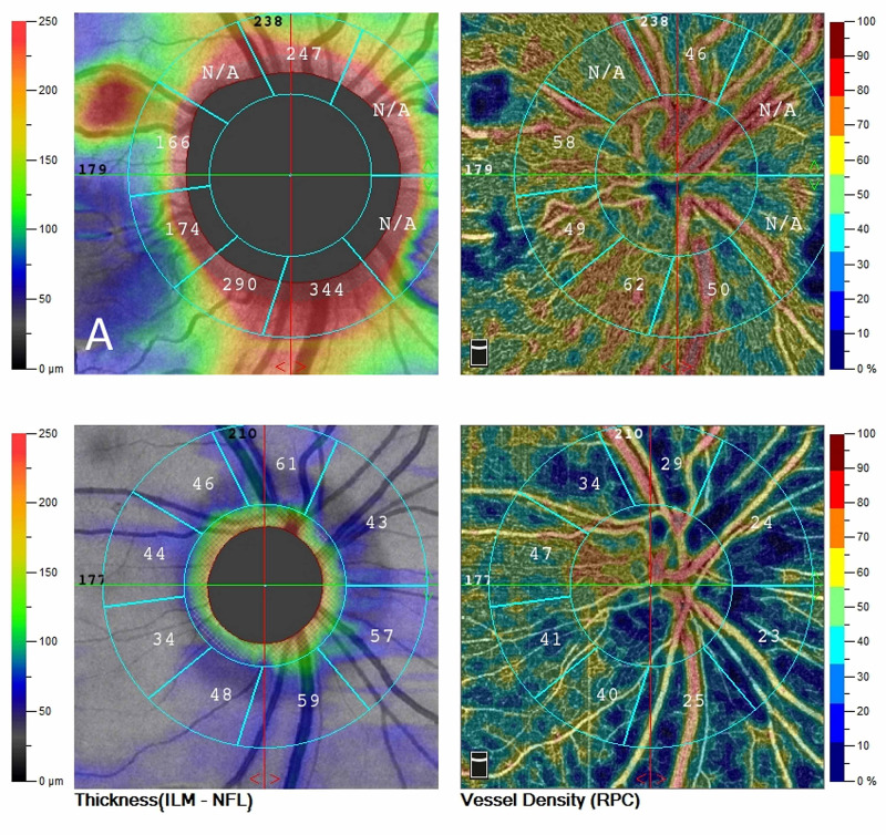
Figure 2. OCT angiography of the optic disc.
A. Upon presentation, diffuse edema with normal vascular perfusion was observed. B. A month later, optic disc atrophy was established with associated thinning of the peripapillary fiber layer. Extensive areas of reduced perfusion are observed in the peripapillary area. Flow remains within normal limits temporal to the optic disc.
OCT: optical coherence tomography

