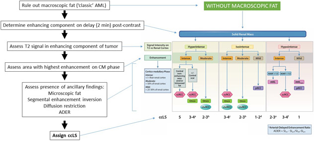
Figure 2:
Clear Cell Likelihood Score (ccLS) diagnostic algorithm and image interpretation. The ccLS is a 5-tier classification that denotes the likelihood of a mass representing a clear cell carcinoma: 1, very unlikely; 2, unlikely; 3, equivocal; 4: likely; and 5, highly likely. First, reviewer must exclude the presence of macroscopic fat (i.e. ccLS is not applicable to masses with macroscopic fat), which would be diagnostic of angiomyolipoma. Second, assessment of delayed post-contrast images (2 minutes) helps define the enhancing portions of the mass. Renal mass signal characteristics and enhancement in subsequent steps is evaluated in the enhancing portion of the mass only (i.e. avoid non-enhancing portions, cystic degeneration, etc.). Third, the mass is classified as hyperintense, isointense, or hypointense relative to the renal cortex based on T2-weighted images (preferable on single-shot fast spin echo images without fat suppression). Fourth, renal mass is classified based on presence of intense (similar or higher), moderate (approximately 50%), or mild (approximately 25-30%) enhancement during the corticomedullary phase relative to the enhancement in the ipsilateral renal cortex. Assessment of enhancement is made with a region of interest (approximately 100 mm2) placed in the area of the tumor that demonstrates the most marked contrast enhancement during the corticomedullary phase on the basis of a visual assessment. Next, the mass is further characterized based on the presence of microscopic fat, segmental enhancement inversion, diffusion restriction (i.e. higher and lower signal intensity than renal parenchyma on b800 DWI and ADC, respectively), and the arterial-delayed enhancement ratio (ADER). Lastly, a ccLS is assigned. a – ccLS = 3 if segmental enhancement inversion (SEI) is present; b – ccLS = 2 if SEI is present; c – ccLS = 4 if microscopic fat is present; d – ccLS = 2 if enhancement is between 25% and 50%; e – ccLS = 2 if homogeneous or marked restriction on diffusion weighted images (DWI); f – ccLS = 3 if homogeneous or marked restriction on DWI, ccLS =4 if heterogeneous. AML = angiomyolipoma, CM = cortico-medullary, ccRCC = clear cell renal cell carcinoma, Onco = oncocytoma, chrRCC = chromophobe renal cell carcinoma, pRCC = papillary renal cell carcinoma, SIart = arterial phase signal intensity (SI), SIpre = pre-contrast SI, SIdel = delayed phase SI. (Modified with permission from [22])

