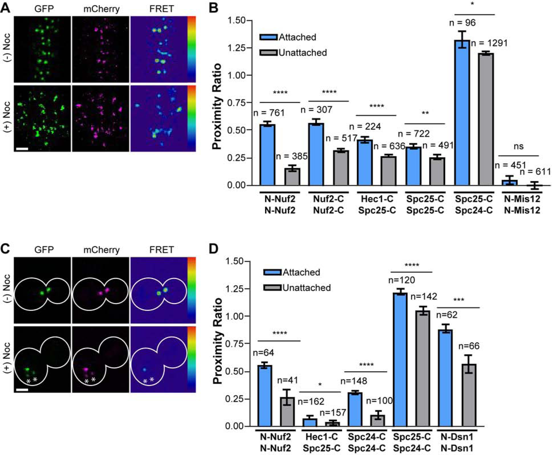
Figure 4. Microtubule attachment clusters Ndc80C in both human and budding yeast kinetochores.

A) Micrographs of mitotic HeLa cells expressing N-Nuf2/N-Nuf2 with and without nocodazole (Noc) treatment. B) Nocodazole treatment reduces FRET between Ndc80C subunits. Measurements are from metaphase (blue) and nocodazole-treated (gray). C) Micrographs of budding yeast metaphase cells expressing N-Nuf2/N-Nuf2, with or without nocodazole. Asterisks highlight clusters of unattached kinetochores. D) Same as in B) but for budding yeast kinetochores. For A) and C), FRET micrographs are scaled equivalently; GFP and mCherry micrographs are scaled for ease of viewing. Scale bar, 2 μm. For D) and B), bars are average proximity ratio ± SEM. The number of measurements is indicated above the bars. All data are from N ≥ 3 experiments. Statistical significance was evaluated using the Mann Whitney test, ns = not significant; * = p<0.05; ** = p<0.01; *** = p<0.001; **** = p<0.0001. See also Figure S5 and Table S1.
