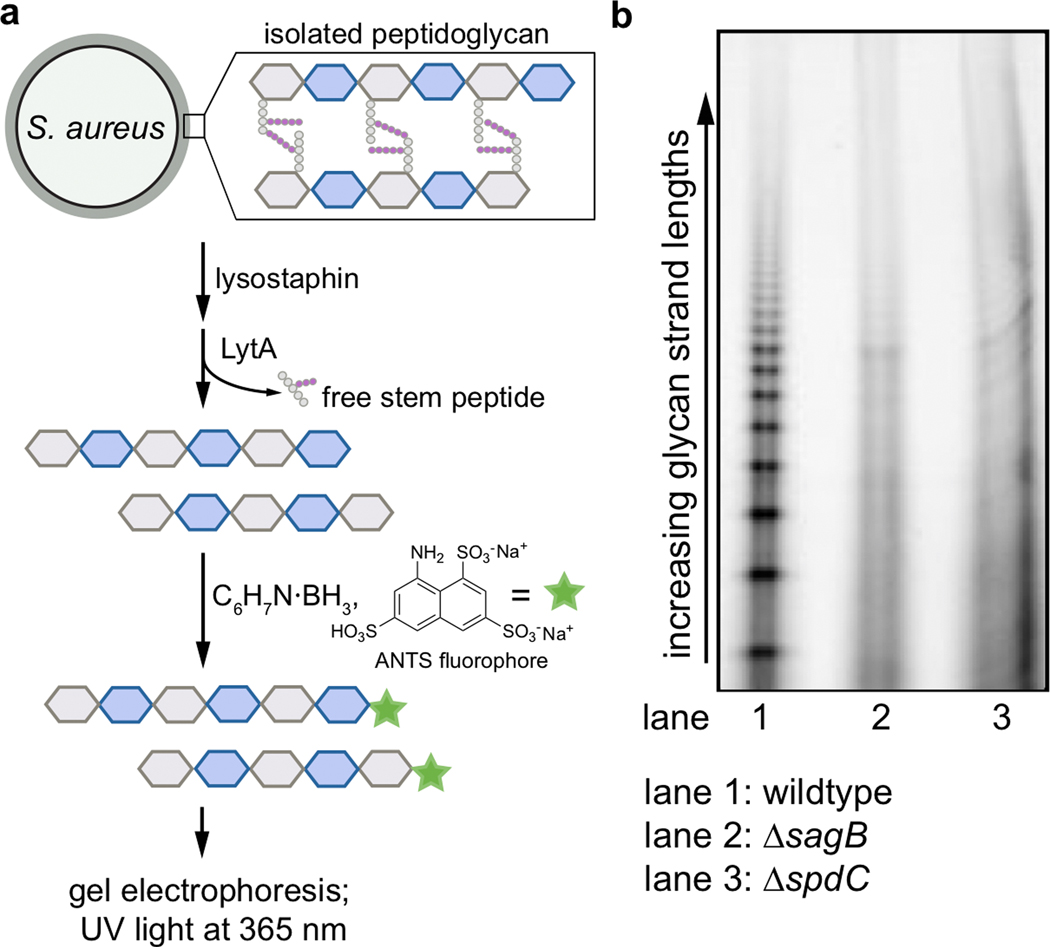
Extended Data Fig. 4. Glycan strand lengths are longer in the absence of spdC or sagB.
a, Sacculi were isolated from wild-type, ΔsagB, and ΔspdC S. aureus strains. The purified peptidoglycan was then treated with the endopeptidase lysostaphin, which cleaves the PG crosslinks, and the amidase LytA, which removes the remaining stem peptides. These denuded glycan strands were then labeled at the reducing end by reductive amination with the anionic fluorophore 8-aminonaphthalene-1,3,6-trisulfonic acid (ANTS), and separated by gel electrophoresis for in-gel imaging (UV excitation at 365 nm, visible emission). b, In lane 1, showing PG isolated from a wild-type strain, shorter glycan strands are visualized as a discrete ladder. In lanes 2 and 3, representing glycan strands of ΔsagB and ΔspdC respectively, this discrete ladder of short glycan strands is lost.

