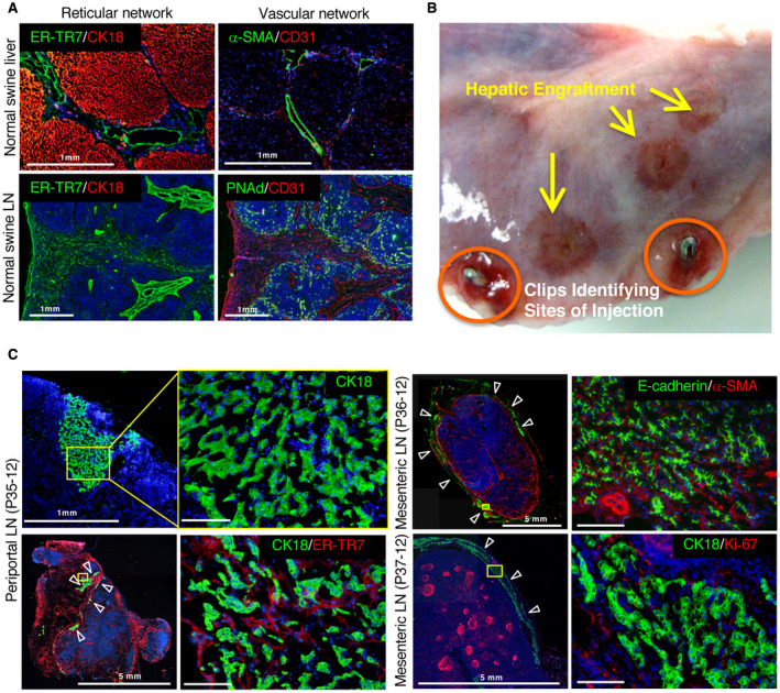
Fig. 2.

Liver tissue generated by hepatocyte transplantation in LNs of pigs undergoing RPVL and partial hepatectomy. (A) Reticular and vascular networks in control pig liver and LN. Normal pig native liver immunostained for hepatocytes with (top left) CK18 (red) and for reticular fibroblasts and fibers (ER‐TR7, green) and (top right) endothelial cells (CD31, red) or hepatic artery/PV with α‐SMA (green). Normal pig LN stained with (bottom left) CK18 (red) and ER‐TR7 (green) or (bottom right) CD31 (red) and high endothelial venules (PNAd, green). (B) Macroscopic view of a transplanted LN with surgical clips (circled in orange) to identify the sites where the hepatocytes were injected. Additional hepatic engraftments are observed on the surface of the LN and are indicated by yellow arrows. (C) Immunostaining of the engrafted hepatocytes in LN from transplanted animals P35‐12, P36‐12, and P37‐12. The yellow rectangles highlight the area of magnification presented on the right side of the panel. (left) P35‐12: general view of the injected LN and the limited engraftment of hepatocytes (CK18, green). White arrowheads indicate hepatocyte engraftment in a LN. (right) General view and higher magnification of the injected LN for P36‐12 (top) and P37‐12 (bottom). E‐cadherin and CK18 (green) stained the hepatocytes; α‐SMA (red) stained the vasculature; and Ki‐67 (red) stained the proliferating cells. All immunostainings were counterstained with Hoechst 33342 dye. Scale bar, 100 μm or as indicated.
