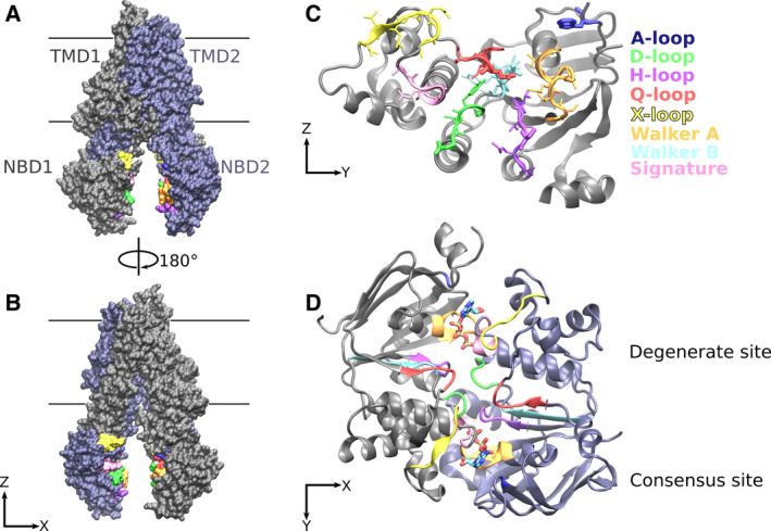
Fig. 1.

ABC transporter structure. (A, B) Side views of inward‐facing ABCC7 (PDB ID: 5UAK [70]) showing the TMD1 and NBD1 in gray, while TMD2 and NBD2 are in violet. The NBD motifs are colored as labeled in panel C. (C) The NBD motifs are highlighted by color and their side chains shown as sticks. The NBD is oriented to show the NBD surface that constitutes the NBD‐NBD interface. (D) Nucleotide‐bound ABCC7 (PDB ID: 6MSM [68]) structure is used to visualize the closed NBD dimer. The NBDs are seen from the TMD domain. For simplicity, the TMD is not shown. All noncanonical motifs in the NBD1 and in NBD2 are clustered at the degenerate NBS1.
