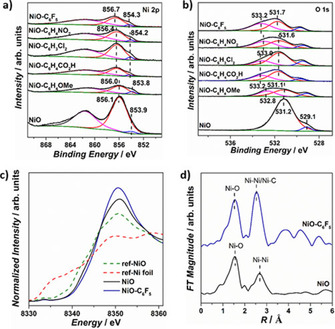
Figure 3.

High‐resolution core XPS spectra of a) Ni 2p and b) O 1s of bare NiO and all modified NiO samples. c) Normalized Ni K‐edge XANES spectra of NiO, NiO−C6F5, reference Ni foil, and reference NiO samples. d) Fourier transformations of k 3‐weighted EXAFS spectra of bare NiO and NiO−C6F5 samples.
