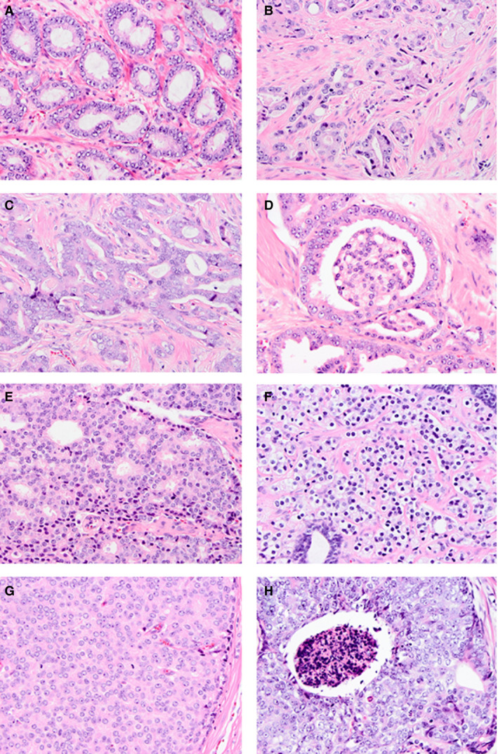
Figure 1.

Overview of Gleason growth patterns. A, Gleason pattern 3 well‐delineated glands. B–E, Gleason pattern 4 poorly formed (B), fused (C), glomeruloid (D) and cribriform (E) architecture. F–H, Gleason pattern 5 single cells/cords (F), solid sheets (G), and comedonecrosis (H). Haematoxylin and eosin.
