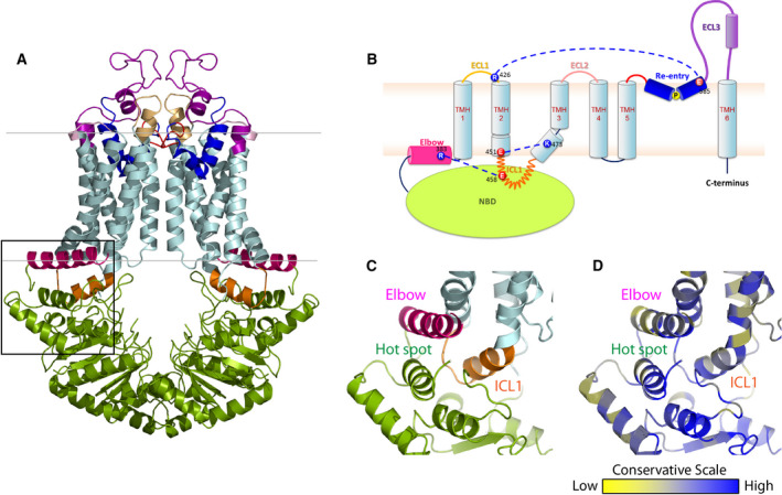
Fig. 1.

The structural organization of human ABCG2. (A) Inward‐facing configuration of the homodimeric ABCG2 transporter in the ATP‐free state (PDB ID: 6ETI). The black box delimits the THB cluster of three conserved helices at the transmission interface, NBD (green), elbow helix (pink), and first ICL (orange). Transmembrane‐spanning helices (light blue), extracellular loop 1 (yellow), re‐entry helix (dark blue), extracellular loop 3 (purple). (B) Membrane topology of ABCG2 half transporter indicating the salt bridge interactions at both membrane interfaces (blue dotted lines). Colors are as in panel (A). (C) Zoom‐in the black box of (A) shows adjacent structural arrangement of three important domains: NBD, elbow helix, and ICL1 at transmission interface. (D) The conservation analysis of (C) is given as color‐gradient: low conservation (yellow) to high conservation (blue).
