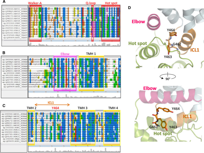
Fig. 2.

Sequence alignment of mammalian ABCGs and the first half of yeast PDRs. Multiple sequence alignment was analyzed using ClustalX2. Conserved residues are highlighted with the conservation scale as the height of gray bars at the bottom of each residue. (A) Conservative regions of Walker A, Q‐loop, and hot spot helix in the NBD are in red boxes. (B) Conserved regions of elbow helix (pink) and transmembrane helix 1(yellow). (C) Conserved regions in transmembrane helices 2, 3, and 4 (yellow). The positions of conserved residues glycine 462, tyrosine 463, and tyrosine 464 of human ABCG2 are marked in the first ICL among ABCGs family proteins. (D) Zoom‐in side views show side chains as sticks of three conserved residues in ICL1, G462, Y463, and Y464, respectively. NBD (green), elbow helix (pink), ICL1 (orange), transmembrane helices (light blue).
