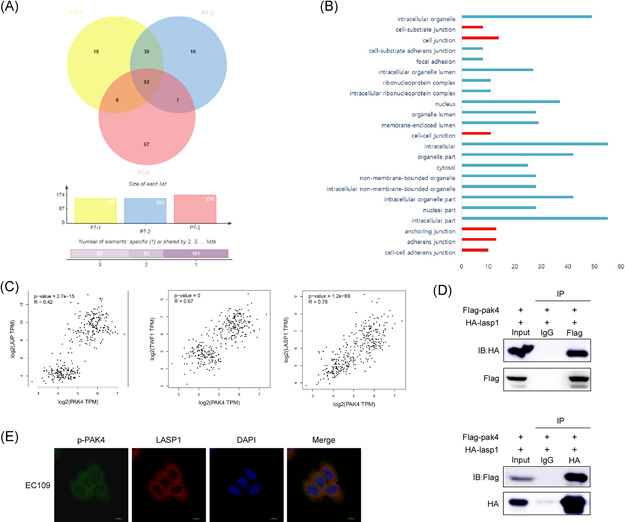
Figure 4.

PAK4 interacts and colocalizes with LASP1. (A) The potential binding proteins of PAK4 were screened by pull‐down assay and mass spectrometry analysis. The Wayne diagram shows the 38 potential binding proteins. (B) Gene ontology functional analysis of 38 genes was performed. (C) The correlation between the expression of PAK4 and JUP, TWF1 and LASP1 in esophageal carcinoma and esophageal normal tissues was analyzed by the GEPIA database. Statistical analysis was performed using Spearman's nonparametric correlation test. (D) PAK4 binds with LASP1 in 293T cells. The pcDNA3.1‐Flag‐pak4 and pcDNA3.1‐HA‐LASP1 plasmids were transiently transfected into 293T cells, Flag‐PAK4 was immunoprecipitated by anti‐Flag, and coimmunoprecipitated HA‐LASP1 was detected by anti‐HA (top). Conversely, HA‐LASP1 was immunoprecipitated by anti‐HA, and coimmunoprecipitated Flag‐PAK4 was detected by anti‐Flag (bottom). (E) p‐PAK4 and LASP1 colocalize in EC109 cells. p‐PAK4 and LASP1 were detected by immunofluorescence staining with p‐PAK4 and LASP1 primary antibodies, a FITC‐conjugated goat anti‐mouse secondary antibody for p‐PAK4, and a TRITC‐conjugated goat anti‐rabbit antibody for detection of LASP1. The data shown are representative of results from triplicate independent experiments. IB, immunoblot; IP, immunoprecipitation [Color figure can be viewed at wileyonlinelibrary.com]
