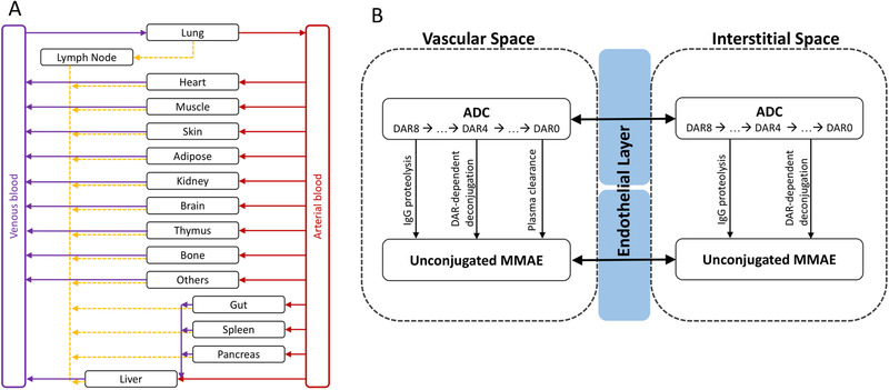
Figure 4.

PBPK model of a vc‐MMAE antibody‐drug conjugate. (A) Structure of PBPK model at whole‐body level. Organs are represented by black rectangle and connected by blood flow (red and purple lines) and lymphatic flow (orange dashed line). (B) Structure of PBPK model at tissue level. Each tissue consists of vascular space, endothelial layer, and interstitial space. The distribution of antibody‐drug conjugate and unconjugated MMAE between vascular and interstitial space through convection, diffusion, or transcytosis is simplified and represented by the black double arrow. The formation of unconjugated MMAE from an antibody‐drug conjugate is linked by IgG proteolysis, drug‐to‐antibody ratio‐dependent deconjugation, and drug‐to‐antibody ratio‐dependent plasma clearance (vascular space only).
