
Figure 4.
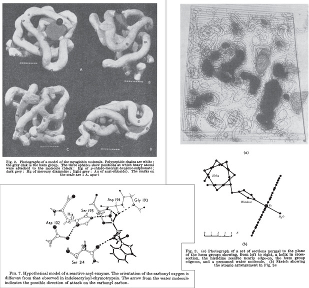
Top left: Clay model of the first X‐ray structure of a protein, myoglobin, at 6 Å resolution. [26a] Right: electron density sections of myoglobin at 2 Å resolution and sketch of groups coordinated to iron. [26b] Bottom left: Model of the catalytic triad and oxyanion hole of chymotrypsin, as inferred from crystal‐structures. [31c]