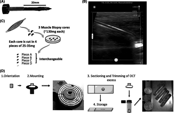
FIGURE 1.

Overview of the core needle biopsy procedure, biopsy handling, and storage. Biopsies were obtained from different muscles including upper (BB) or lower limbs (vastus lateralis, tibialis anterior [TA], and gastrocnemius). Each pass of the 10‐gauge needle obtains a piece of muscle about 3 mm in diameter and about 15 mm in length; image is of a muscle core prior to removal of the muscle core from the needle chamber, A. A handheld ultrasound system is used prior to all procedures to ensure to avoid blood vessels and nerves; ultrasound static image shows about 3 cm insertion of needle into TA at maximal insertion prior to vacuum‐assisted coring, B. 21 Each muscle core is dissected into 25 to 35 mg pieces, each of which can be used for either histologic assessments, protein analyses, RNA analyses, and we thus consider “interchangeable,” C. All small pieces are stored in liquid nitrogen (not depicted). Each piece is preserved in liquid nitrogen until sectioning. Once retrieve from liquid nitrogen, the muscle is mounted directly on the chuck. After 10 μm thick sectioning onto glass slides, we remove the excess OCT such that the remaining muscle can be replaced in the cryovial for long‐term storage in liquid nitrogen. Sections on slides are stored in −80°C, D
