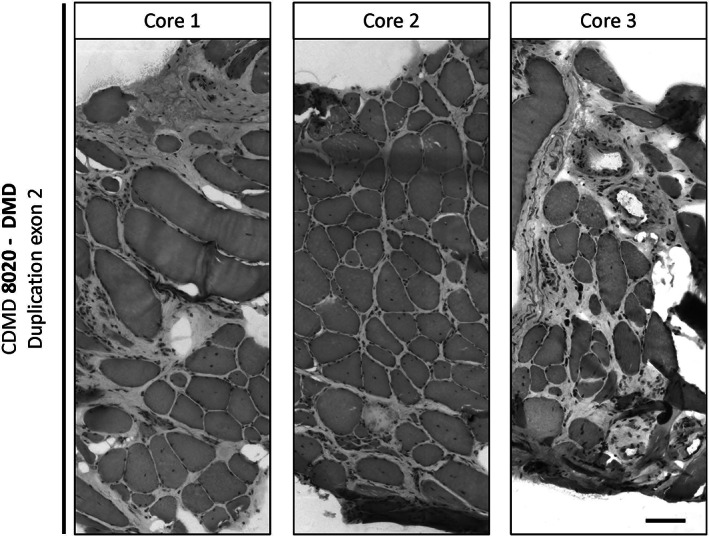
FIGURE 3.

Heterogeneity of pathology across and within different muscle cores taken from the same muscle. Three cores were taken from the vastus lateralis of patient CDMD 8020 (DMD duplication exon 2) and were all frozen using the “Cassette in LN2 direct” method. Each piece was trimmed for about 100 μM depth of muscle tissue before sampling serial 10 μM sections and subsequently stained with H&E. Images were assembled as a mosaic at 20× to reveal well‐preserved structures and myofibers regardless of the degree of fibro‐fatty involvement. Scale bar is 100 μM
