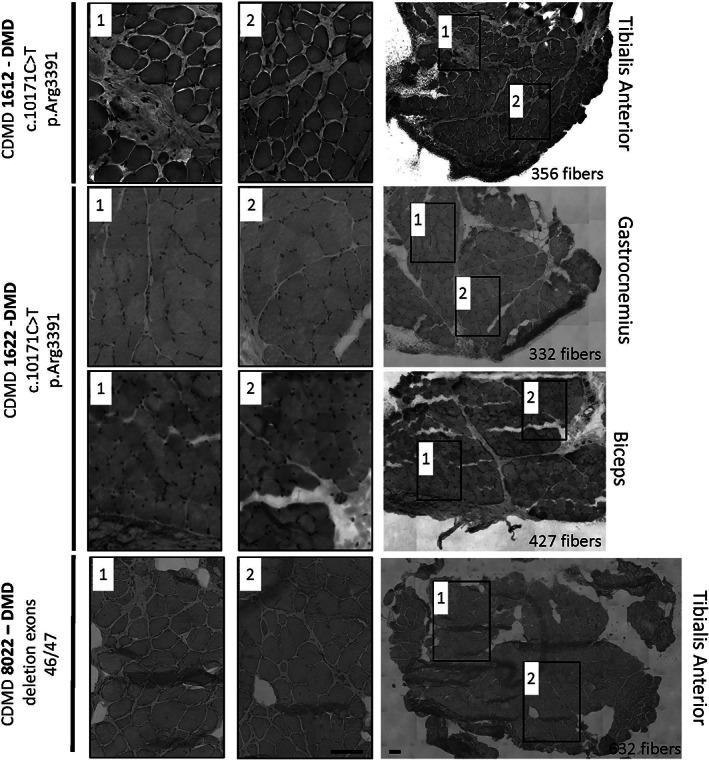
FIGURE 4.

Needle muscle biopsy and “Cassette in LN2” tissue processing provides sufficient muscle for histological analysis of dystrophic and healthy muscle. Core needle biopsies processed using the “Cassette in LN2 direct” method were analyzed for histology quality and number of myofibers in different muscles: CDMD1612 (DMD c.10171C>T, p.Arg3391*) was sampled from the biceps brachii at age 6 y, CDMD1622 (DMD c.10402G>T, p.Glu3468*) was sampled from gastrocnemius at age 6 y, and CDMD8022 (DMD out of frame deletion of exons 46‐47) was sampled from tibialis anterior at age 21 y. Each piece was trimmed for about 100 μM before sampling a 10 μM section for H&E. The whole cross‐section (right panel) was counted for myofibers and the number of fibers is indicated for each cross‐section in the lower right corner of the image. Two higher magnification images (first and second images from the left) from each whole cross‐section demonstrate lack of freeze artifact or distortions in muscles with varying fibro‐fatty replacement. Images for whole cross‐sections were reconstructed as a mosaic at 20×. Scale bar is 100 μM
