FIGURE 4.

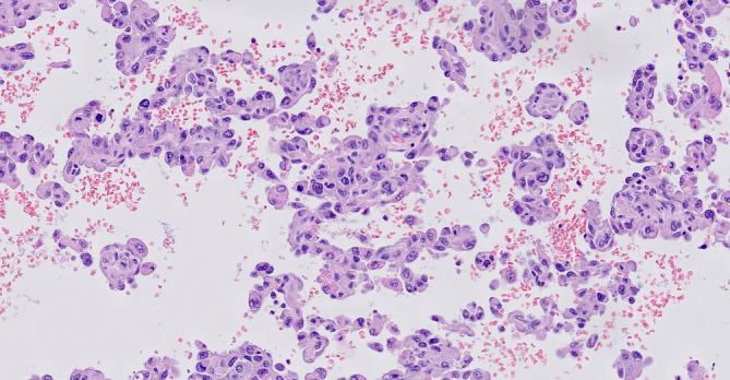
Detail of the angiosarcomatous component. Highly atypical endothelial cells can be seen and mitotic figures are present. The vascular spaces contain erythrocytes (hematoxylin and eosin ×200)

Detail of the angiosarcomatous component. Highly atypical endothelial cells can be seen and mitotic figures are present. The vascular spaces contain erythrocytes (hematoxylin and eosin ×200)