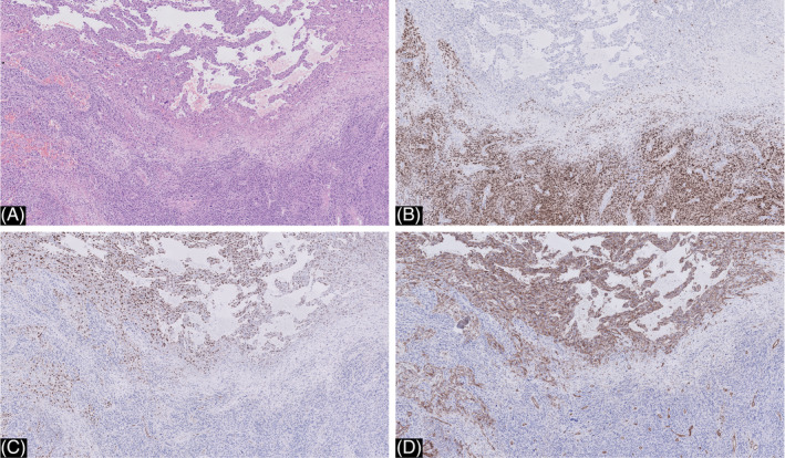
FIGURE 5.

A, close‐up of the melanoma metastasis in the inguinal lymph node showing both components (hematoxylin and eosin ×20). B, immunohistochemistry for SOX‐10 was positive in the conventional melanoma and completely negative in the vascular component (SOX10 ×20). C and D, immunohistochemistry for Erg (C) and CD 31 (D) was positive in the vascular component and completely negative in the conventional melanoma (ERG ×20)
