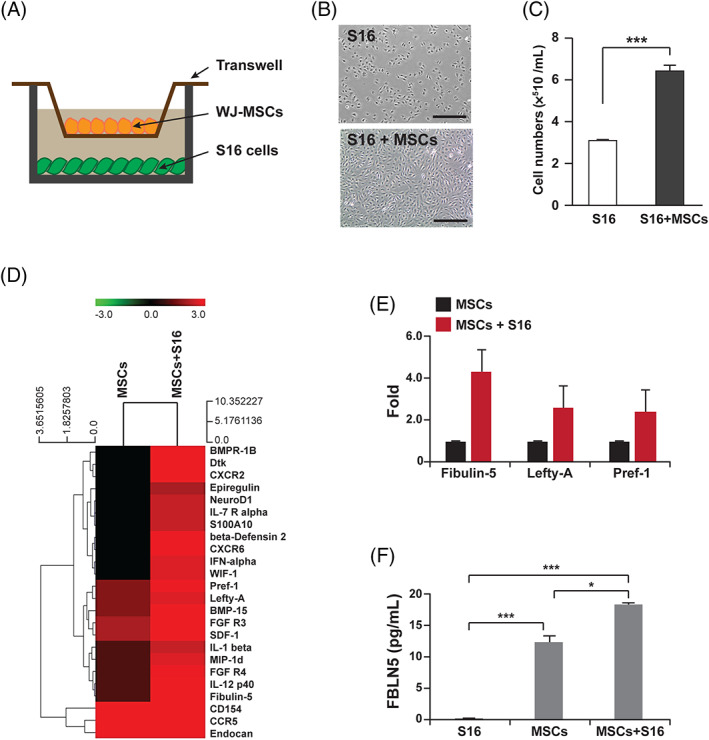
FIGURE 1.

Identification of WJ‐MSCs derived paracrine factors affecting Schwann cell proliferation. A, A schematic diagram for the co‐cultivation system of MSCs and S16 cells. B, Images of S16 cells after 24 hours of cultivation with or without WJ‐MSCs. Scale bars, 400 μm. C, Quantification of total number of S16 cells counted at each indicated condition. Statistical significance was determined using the unpaired Student's t‐test with Welch's correction (***P < .001). D, A heatmap comparing the expression of secreted proteins in the media collected from the indicated culture conditions. E, Quantifications of fold changes in spot intensities for Fibulin 5, Lefty‐A, and Pref‐1. F, The concentration of FBLN5 contained in the culture media collected from each indicated condition was measured using ELISA. Statistical significance was determined using one‐way ANOVA followed by Tukey's post hoc test (*P < .005, ***P < .001)
