FIGURE 2.

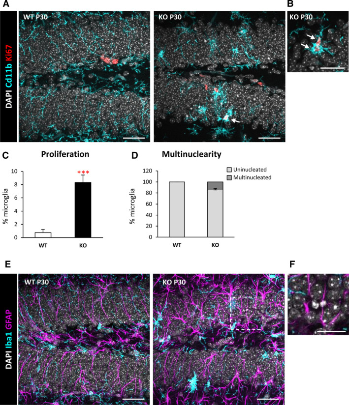
Increased proliferation and multinuclearity in the dentate gyrus (DG) of postnatal day 30 (P30) Cstb knockout (KO) mice. A, DG general view of both wild‐type (WT) and Cstb KO P30 mice. Proliferating cells were stained for Ki67 (marker for all the active phases of the cell cycle), nuclei (4,6‐diamidino‐2‐phenylindole [DAPI]), and microglia (CD11b). B, High‐magnification image of proliferating Ki67+ microglia in P30 Cstb KO mice. Arrows in A and B point to Ki67+ microglia. C, Percentage of proliferating microglia assessed by the marker Ki67. D, Proportion (in %) between uninucleated and multinucleated microglia in the septal hippocampus of WT and Cstb KO P30 mice. E, Representative images of the DG in WT and Cstb KO mice stained for DAPI (nuclei), microglia (Iba1), and astrocytes (glial fibrillary acidic protein [GFAP]). F, High‐magnification image of an apoptotic cell not phagocytosed by either microglia or astrocytes in Cstb KO mice. Bars represent the mean ± standard error of the mean. ***P < .001 by one‐tailed Student t test. Scale bars = 40 µm (A), 20 µm (B), 40 µm (E), 20 µm (F); z‐thickness = 7 μm (A, E), 3.5 μm (B)
