
FIGURE 8.
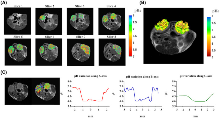
A, Two‐dimensional multislice tumor extracellular pH (pHe) map for a breast‐tumor murine model. B, Three‐dimensional pH map rendering. C, Calculated pH gradients along the three main axes inside the left tumor region showing T2‐weighted image, overimposed pH map, and pH gradients along the A‐axis (red), B‐axis (blue), and C‐axis (green).