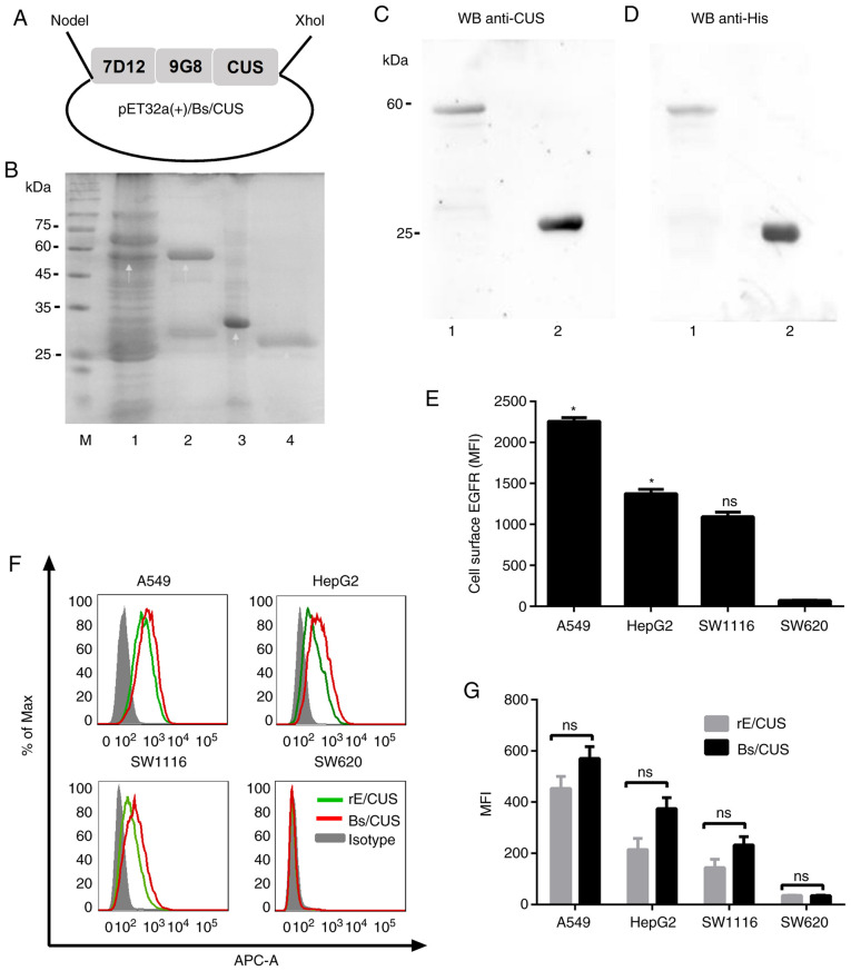
Figure 1.
Expression and characterization of RIT. The pET32a(+)/Bs/CUS plasmid was transformed into E. coli BL21 (DE3) cells. (A) Schematic structure of pET32a(+)/Bs/CUS. (B) Use of 12% SDS-PAGE under reduced conditions. M, protein marker; 1, E. coli BL21 (DE3) transformed with pET32a(+)/Bs/CUS induced by 1 mM IPTG; 2, Bs/CUS purified by Ni-NTA column. 3, 7D12-9G8; 4, CUS. (C and D) Western blot confirmed the expression of Bs/CUS and CUS by using the primary antibodies against CUS and His tag, respectively. 1, Bs/CUS; 2, CUS. (E) EGFR expression on different cell lines. (F and G) Affinity of Bs/CUS compared with that of rE/CUS by flow cytometry. MFI, mean fluorescence intensity. *P<0.05 vs. SW620.

