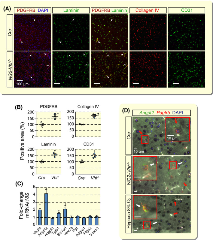
FIGURE 1.

Vhl inactivation in NG2 cells results in neurovascular remodelling and expansion. A, Representative immunofluorescence (IF) images of frozen brain sections from Cre− control and NG2‐Vhl−/− mice (age 9‐10 wk) stained for markers of pericytes (PDGFRB), basement membrane (laminin and collagen IV) and endothelial cells (CD31); cortex; scale bar, 100 µm. Nuclei were stained with 4′,6‐diamidino‐2‐phenylindole (DAPI, blue fluorescence). There is complete overlap between PDGRFB and laminin IF staining. White arrows exemplify double‐positive structures. B, Quantification of PDGFRB‐, laminin‐, collagen IV‐ and CD31‐positive area expressed as percentage of Cre− control, (n = 3). C, Relative mRNA transcript levels in NG2‐Vhl−/− cortex expressed as fold‐change compared with Cre− control, (n = 3‐5). D, Representative images of multiplex RNA FISH of cortical tissue from Cre− control, NG2‐Vhl−/− and hypoxic wild‐type mice (normobaric hypoxia, 8% O2 for 24 h) detecting angiopoietin 2 (green fluorescent signal) and Pdgfrb‐expressing cells (red fluorescent signal). White arrows depict cells positive for both Angpt2 and Pdgfrb; red arrows depict cells in the direct vicinity of micro‐vessels that are positive for Angpt2 and negative for Pdgfrb. White structures represent auto‐fluorescent red blood cells. Nuclei were stained with DAPI (blue fluorescent signal); scale bar, 10 µm. Data are represented as mean ± SEM; two‐tailed Student's t test; *P < .05, † P < .01 and ‡ P < .001 compared with Cre− control. Adgre1, adhesion G‐protein‐coupled receptor E1; Angpt1, angiopoietin 1; Angpt2, angiopoietin 2; Pgf, placental growth factor; Ptgs2, prostaglandin‐endoperoxide synthase 2; Slc7a5, solute carrier 7a5; Tgfb1, transforming growth factor beta 1; Vcam1, vascular cell adhesion molecule 1; Vegfa, vascular endothelial growth factor A; Wnt7b, wingless‐type MMTV integration site family, member 7B
