FIGURE 2.

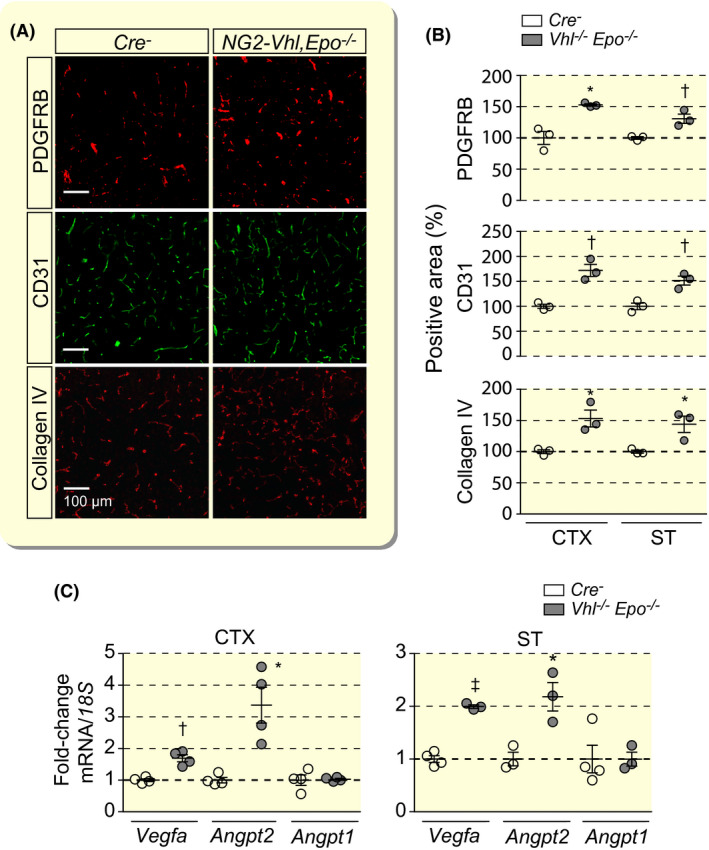
Cerebral vascular expansion in NG2‐Vhl−/− mutant mice is not EPO‐dependent. A, Representative immunofluorescence images of frozen brain sections from Cre− control and NG2‐Vhl−/−Epo−/− (Vhl‐/‐Epo−/−) mice stained for markers of pericytes (PDGFRB), endothelial cells (CD31) and basement membrane (collagen IV); shown is cortex; scale bar, 100 µm. B, Quantification of cortical and striatal PDGFRB‐, CD31‐ and collagen IV‐positive areas expressed as percentage of Cre− control, (n = 3). C, Relative mRNA levels in cerebral cortex (CTX) and striatum (ST) from NG2‐Vhl−/−Epo−/− mice expressed as fold‐change compared with Cre− control, (n = 4). Data are represented as mean ± SEM; two‐tailed Student's t test; *P < .05, † P < .01 and ‡ P < .001. Angpt1, angiopoietin 1; Angpt2, angiopoietin 2; Vegfa, vascular endothelial growth factor A
