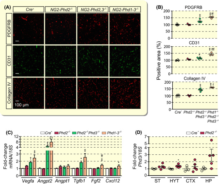
FIGURE 3.

Combined inactivation of Phd2 and Phd3 but not inactivation of Phd2 alone promotes cerebral vascular expansion. A, Representative immunofluorescent images of frozen brain sections from Cre− control, NG2‐Phd2−/−, NG2‐Phd2−/−Phd3−/− (NG2‐Phd2,3−/−) and NG2‐Phd1−/−Phd2−/−Phd3−/− (NG2‐Phd1‐3−/−) mice stained for PDGFRB, CD31 and collagen IV; scale bar, 100 µm. B, Quantification of cortical PDGFRB‐, CD31‐ and collagen IV‐positive areas expressed as percentage of Cre− control (n = 3‐6). C, Relative cortical mRNA transcript levels in homogenates from cortex of Cre− control, NG2‐Phd2−/−, NG2‐Phd2−/−Phd3−/− and NG2‐Phd1−/−Phd2−/−Phd3−/− mice expressed as fold‐change compared to Cre− control (n = 4‐11). D, Phd3 transcript levels in striatum (ST), hypothalamus (HYT); cortex (CTX) and hippocampus (HP) from Cre− control and NG2‐Phd2−/− mice. Data are represented as mean ± SEM; one‐way ANOVA followed by Tukey's post hoc analysis; *P < .05, † P < .01 and ‡ P < .001 compared with Cre− control; f P < 0.05, ff P < 0.01, and fffP < 0.001 compared with NG2‐Phd2−/−Phd3−/− mice. Angpt1, angiopoietin 1; Angpt2, angiopoietin 2; Cxcl12, C‐X‐C motif chemokine 12; Fgf2, fibroblast growth factor 2; Tgfb1, transforming growth factor beta 1; Vegfa, vascular endothelial growth factor A
