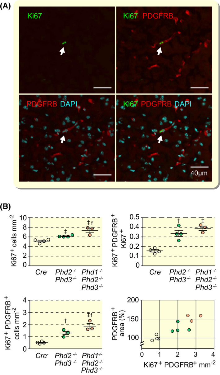
FIGURE 4.

Combined inactivation of Phd2 and Phd3 in NG2 cells promotes pericyte proliferation. A, Representative high‐power immunofluorescence images of proliferating pericytes in cerebral cortex depicted by white arrows. PDGFRB‐positive cells are identified by red fluorescence and proliferating cells by Ki67 staining (green fluorescent signal). Nuclei were stained with 4′,6‐diamidino‐2‐phenylindole (DAPI, blue fluorescence); scale bar, 40 µm. B, Quantification of cells in cerebral cortex from Cre− control, NG2‐Phd2−/−Phd3−/− and NG2‐Phd1−/−Phd2−/−Phd3−/− mice; (age 9‐10 wk, n = 3‐4). Left upper panel, Ki67+ cells; left lower panel, proliferating pericytes (Ki67+PDGFRB+); right upper panel, ratio of Ki67+PDGFRB+ cells over total number of Ki67+ cells; right lower panel, correlation between pericyte covered area (PDGFRB+ cells) and proliferating pericytes; Cre− control (empty circles); NG2‐Phd2−/−Phd3−/− (green circles); NG2‐Phd1−/−Phd2−/−Phd3−/− mice (coral‐colored circles); Pearson's r = 0.8895, P = .0002 (two‐tailed). Data are represented as mean ± SEM; one‐way ANOVA followed by Tukey's post hoc analysis, † P < .01 and ‡ P < .001 compared with Cre− controls. f P < 0.05 compared with NG2‐Phd2−/−Phd3−/− mice
