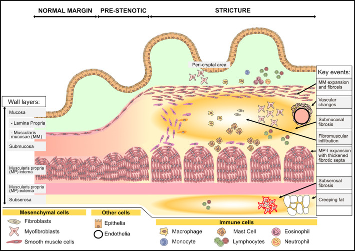
Figure 1.

An overview of strictured tissue showing changes in intestinal wall layers and cell populations. A schematic of the most apparent histological changes in the intestinal wall during the progression from normal to strictured tissue is shown. This includes increased intestinal wall thickness due to expanded smooth muscle cells, and hence muscle layers, concomitant with fibrotic changes in the submucosa and subserosa. Histological features described in the text are highlighted in the right column as ‘Key events’ with the location of the event indicated by an arrow. See the text and subsequent figures for details
