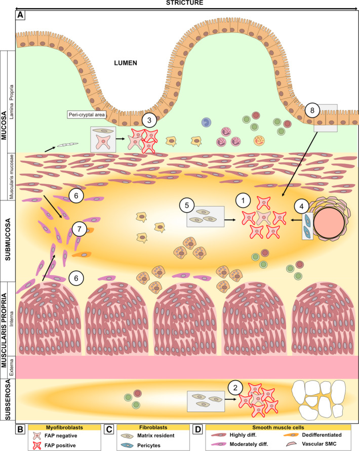
Figure 3.

Schematic of the strictured wall with a focus on location of mesenchymal cells. A, The transition into strictured tissue involves an increase in extracellular matrix deposition, fibrosis (indicated by yellow/orange) particularly in the submucosa and subserosa, and expansion of the muscle layers along with infiltration of cells. There is accumulation of predominantly FAP+ myofibroblast in the layers most affected by fibrosis, namely, in ① the submucosa and ② subserosa. However, similar enrichment is also observed in the ③ peri‐cryptal area of the mucosa. Grey boxes show the condition in normal (non‐strictured) tissue with arrows showing the changes that occur inside strictured tissue. The accumulated myofibroblasts might have arisen from ④ pericytes, ⑤ matrix‐resident fibroblasts, or ⑧ epithelial cells (through epithelial‐to‐mesenchymal transition). Also depicted is ⑥ phenotypically changed SMCs along the border between the submucosa and the muscle layers and in the ⑦ fibromuscular infiltration in submucosa. (B‐D) shows the subtypes of the mesenchymal cells depicted in (A), as discussed. See the text for further details
