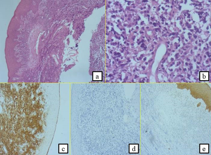
Figure 3.
histological and immunohistochemical examinations leading to the diagnosis of large B-cell lymphoma; presence of large tumour cells having clear and abundant cytoplasm with nuclei showing several marked atypias (A,B). The mitoses and the apoptotic bodies were quite numerous; the tumour was the site of important necrotic changes, the tumour cells strongly expressed CD20 (C); they did not express CD3 (D) and CK (E)

