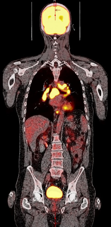
Figure 1.

Follow-up positron emission tomography/CT scan after six cycles showing possible marked disease progression with diffuse fluorodeoxyglucose-avid adenopathy of the mediastinum, bilateral hilar and periaortic regions. Not pictured are the clavicular lymph nodes.
