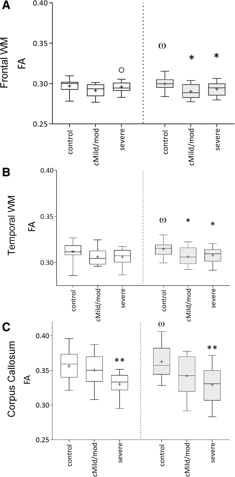
FIG. 2.
Mean regional fractional anisotropy (FA) values in control, complicated mild (cMild)/moderate (mod) traumatic brain injury (TBI), and severe TBI groups showing an interval changes in FA between the initial (3–17 days; white bars) and 12-month (gray bars) imaging time-points. Control subjects showed a significant interval increase in FA in the frontal (A) and temporal (B) white matter, and corpus callosum (C) as expected with normal development (‡p < 0.05). FA was reduced in the frontal and temporal white matter of both the modTBI and severe TBI (sTBI) groups (*p < 0.05) and corpus callosum of the sTBI group (**p < 0.01).

