FIG. 1.
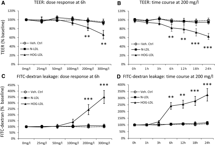
HOG-LDL decreased TEER and induced FITC-dextran leakage in an RPE barrier model. ARPE-19 cells were cultured in transwell plates for 18–21 days to form a confluent monolayer and then treated with SFM for 18 h to achieve quiescence. Cells were then exposed to HOG-LDL or N-LDL at concentrations and time points indicated, versus PBS as a vehicle control. TEER and FITC-dextran leakage were measured; no-treatment and 0-h values served as the baseline, respectively. TEER responses are shown in exposure to (A) HOG- versus N-LDL (0–300 mg/L) at 6 h and (B) HOG- versus N-LDL (200 mg/L) over 0–24 h. FITC-dextran leakage responses are shown in exposure to (C) HOG- versus N-LDL (0–300 mg/L) at 6 h, and (D) HOG- versus N-LDL (200 mg/L) for 0–24 h. Data are presented as percentages relative to the baseline (mean ± SD, n = 3). **P < 0.01 and ***P < 0.001 versus vehicle control. TEER, transepithelial electric resistance; HOG-LDL, heavily oxidized, glycated low-density lipoprotein; SFM, serum-free medium; FITC, fluorescein isothiocyanate.

